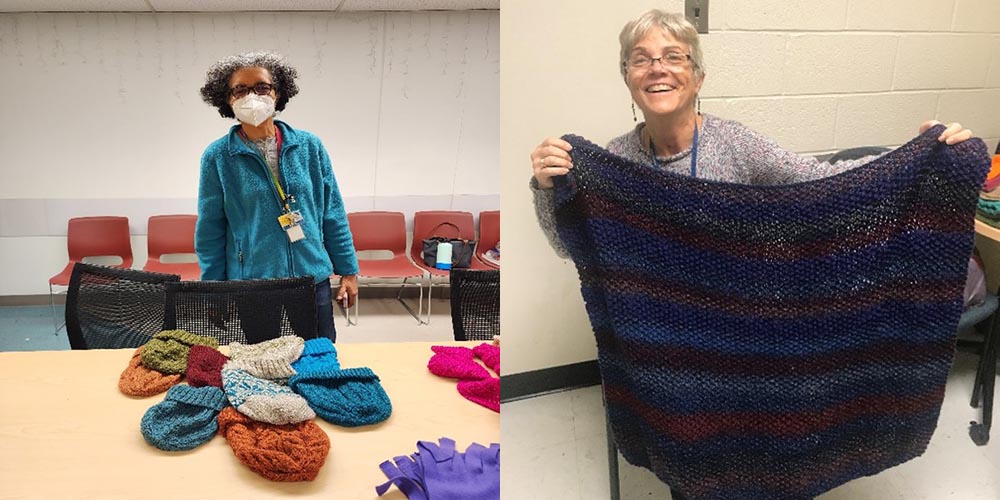
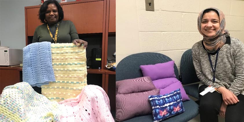
Last year, a few of us in Microbiology who enjoy knitting, crocheting and sewing decided to form a group to provide handmade items that would be useful for patients and their families at Johns Hopkins medical centers.
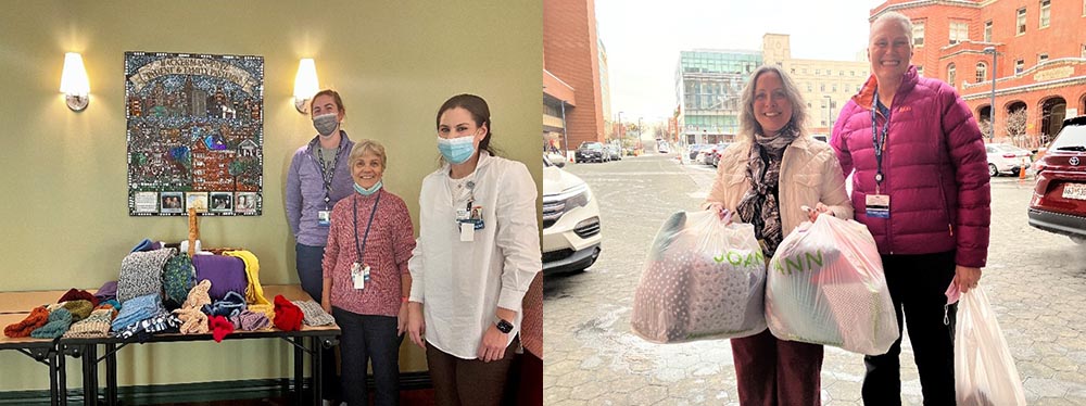
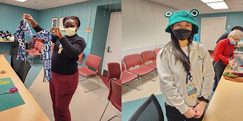
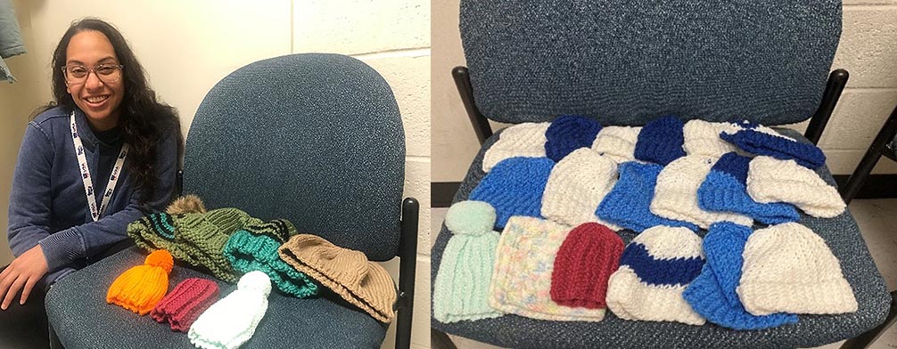
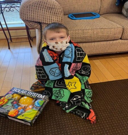
Some of the items include adult and baby hats, scarves, blankets; socks, small pillows, tote bags, and specialty items for the Breast Centers such as drain bags and knitted knockers, and IV and PIC line covers. We serve the Kimmel Cancer center, Bayview Center for addicted adults and their babies, the Breast Centers and the Hopkins House families. Our dedicated group has provided over 180 items since its inception in December 2022!
We would love to have more active members– if you don’t know how to knit or crochet, we can provide basic instruction with supplies (yarn, needles, fabric) and plenty of easy patterns.
Knitting is a great activity, especially during winter months when we don’t get outside as much. It is a great stress reliever, and there’s plenty of winter left-though most items are needed all year long.
Please consider joining our group!
If you’re curious or interested, please contact us by email. We will add you to the newsletter group and let you know when we have periodic meetings.
Last but not least: if you or anyone you know has some craft supplies sitting around the house or stuffed in a closet (fabric, needles, yarn, Velcro, thread, pillow stuffing etc.—we can use anything!) feel free to contact us, we will be happy to take it!
Paula C. Mister, MS, MT, SM(ASCP) and Carrie Holdren-Serrell MS, M(ASCP)
Microbiology Division